
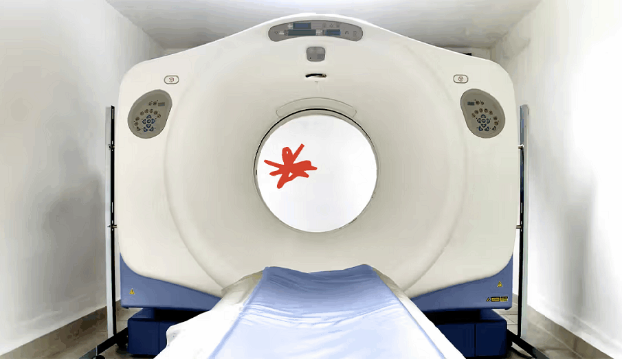
MRI vs. CT Scans: Choosing the Right Tool for Sarcoma Diagnosis
When it comes to diagnosing sarcoma, choosing the right imaging tool is crucial for accurate and effective results. Two commonly used imaging techniques, Magnetic Resonance Imaging (MRI) and Computed Tomography (CT) scans, play significant roles in detecting and evaluating tumors. However, understanding their differences is essential to determine which tool is best suited for the sarcoma diagnosis.
MRI scans utilize strong magnetic fields and radio waves to produce detailed images of soft tissues like muscles and tendons. This non-invasive technique provides high-resolution images that help identify the size, location, and characteristics of sarcoma tumors. On the other hand, CT scans use X-rays and a computer to create cross-sectional images of the body. This imaging technique provides information about bone structure and is more effective in detecting bone metastases and calcifications.
Ultimately, the choice between MRI and CT scans depends on several factors, including the specific type of sarcoma suspected, the location of the tumor, and the patient’s overall health. Consulting with a medical professional is essential to determine the most suitable imaging tool for a timely and accurate sarcoma diagnosis.
Understanding Sarcoma and Its Diagnosis
Sarcoma is a rare type of cancer that originates from connective tissues, such as bone, cartilage, fat, muscle, and blood vessels. Unlike more common cancers like breast or lung cancer, sarcomas make up less than 1% of all adult cancers, which contributes to their being less understood and more challenging to detect and treat. They are generally divided into two categories: soft tissue sarcomas and bone sarcomas, with numerous subtypes under each.
Due to the rarity and complexity of sarcomas, precise diagnostic tools are essential to ensure accurate diagnosis and effective treatment. According to the Sarcoma Oncology Center, early and accurate diagnosis is critical to improving outcomes, as delayed detection can significantly affect prognosis.
Diagnosis usually begins with a medical history review and a physical exam, focusing on symptoms like unexplained lumps, swelling, or persistent pain. Imaging techniques—particularly MRI and CT scans—are crucial in identifying abnormalities and determining tumor characteristics. These imaging modalities help evaluate the tumor’s size, location, and potential spread, which are vital for treatment planning.
Often, a biopsy follows imaging to confirm the diagnosis. The tissue sample provides insight into the tumor’s type and grade, allowing oncologists to tailor treatment approaches, which may include surgery, radiation therapy, or chemotherapy. As highlighted by the Sarcoma Oncology Center, a multidisciplinary team and personalized treatment plans are essential for managing such complex cancers effectively.
The Role of MRI and CT Scans in Sarcoma Diagnosis
MRI and CT scans are fundamental imaging tools in the diagnosis and management of sarcoma, each offering distinct advantages based on the tumor’s location and tissue characteristics.
MRI is particularly effective for evaluating soft tissue sarcomas, thanks to its superior contrast resolution and ability to provide detailed images without ionizing radiation. It allows clinicians to assess the tumor’s size, shape, margins, and relationship to nearby structures such as nerves, vessels, and muscles. Functional MRI techniques can also reveal important information about tumor vascularity and tissue composition, helping to distinguish between benign and malignant masses and guide treatment planning. Gadolinium-based contrast may be used in certain cases to enhance visualization of tumor boundaries and blood supply.
CT scans, on the other hand, are often preferred for detecting bone involvement and metastatic disease, particularly in the lungs. Using X-rays to create cross-sectional images, CT provides excellent detail of bony structures and is commonly used in emergency settings due to its speed and accessibility. While not as detailed as an MRI for soft tissues, CT scans are invaluable for evaluating tumor spread and aiding in staging decisions.
Together, MRI and CT scans complement each other in delivering a comprehensive understanding of sarcoma, improving diagnostic accuracy, treatment planning, and long-term outcomes.
Key Differences Between MRI and CT Scans for Sarcoma Diagnosis
While both MRI and CT scans are invaluable in the diagnosis of sarcoma, they have distinct characteristics that influence their use in clinical practice. The primary difference lies in their imaging techniques; MRI uses magnetic fields and radio waves, while CT scans utilize X-rays. This fundamental difference leads to variations in the types of images produced and the information they convey.
One of the most notable distinctions is the contrast resolution of the images. MRI provides superior soft tissue contrast, making it the preferred choice for visualizing soft tissue sarcomas. In contrast, CT scans excel at visualizing bony structures, making them particularly useful for assessing sarcomas that may invade the bone or have associated bony lesions. Thus, the choice of imaging modality can significantly affect the diagnostic outcome and subsequent treatment plan.
Additionally, the implications of radiation exposure must be considered when choosing between the two modalities. While MRI is a non-ionizing imaging technique, CT scans involve exposure to ionizing radiation. This factor is especially important for younger patients or those requiring multiple imaging studies, as repeated exposure to radiation can increase the risk of secondary malignancies. Therefore, the decision to use one imaging modality over the other may also hinge on the patient’s age and overall health considerations.
Advantages of MRI and CT Scans in Sarcoma Diagnosis
Both MRI and CT scans play a vital role in the early and accurate diagnosis of sarcoma, offering complementary strengths in soft tissue and bone assessment. These imaging tools are central to oncology imaging, providing critical insights that guide treatment planning and surgical decision-making.
MRI is often the preferred imaging modality for evaluating soft tissue sarcomas due to its high-resolution capabilities without the use of ionizing radiation. This makes it particularly valuable for pediatric patients or individuals requiring repeated scans. MRI offers detailed visualization of the tumor’s size, shape, and proximity to surrounding structures—information essential for surgical planning and limb preservation. Additionally, advanced techniques such as diffusion-weighted imaging (DWI) and dynamic contrast-enhanced MRI (DCE-MRI) help assess tumor cellularity, necrosis, and vascularity, providing functional data that can influence treatment decisions.
CT scans, on the other hand, are especially beneficial for detecting bony involvement and metastatic spread, particularly to the lungs and abdomen. CT’s rapid imaging capability is advantageous in emergency settings where time is critical. Its broad availability and cost-effectiveness make it a go-to option for prompt diagnosis. Furthermore, CT’s strength in visualizing both soft tissue and bone makes it a versatile asset in the oncology imaging toolkit.
According to Tellica Imaging, the integration of both MRI and CT imaging in a comprehensive sarcoma workup enhances diagnostic precision and treatment planning. Their team emphasizes that pairing advanced imaging technologies with expert interpretation enables more personalized and effective care for cancer patients.
By leveraging the strengths of both modalities, healthcare providers can achieve a more complete understanding of each patient’s condition—supporting earlier interventions and improving outcomes.
Limitations and Considerations of MRI and CT Scans for Sarcoma Diagnosis
While MRI and CT scans are integral to diagnosing and managing sarcoma, each modality has limitations that must be considered when choosing the appropriate imaging strategy.
MRI excels in soft tissue visualization but has practical challenges. The longer scan duration requires patients to remain still for extended periods, which can be difficult for individuals in pain or those with limited mobility. Motion artifacts may compromise image quality, especially in children or patients with certain medical conditions. Moreover, while MRI provides detailed soft tissue information, it may be less effective in evaluating bone involvement or detecting calcifications—areas where CT offers better clarity.
CT scans, by contrast, are quicker and provide excellent imaging of bone structures and potential metastases, such as in the lungs. However, CT uses ionizing radiation, making repeated scans a concern, particularly for younger patients. CT is also less precise than MRI when it comes to defining soft tissue margins, which can impact surgical planning. Additionally, the use of contrast agents may cause allergic reactions in some individuals, requiring caution or alternative imaging methods.
Ultimately, the choice between MRI and CT—or the decision to combine both—depends on the tumor’s location, suspected spread, and patient-specific factors. In many cases, a multimodal imaging approach is essential for achieving an accurate diagnosis and developing a tailored treatment plan.
Choosing the Right Tool for Sarcoma Diagnosis: MRI vs. CT Scans
When it comes to choosing the most appropriate imaging tool for sarcoma diagnosis, several factors must be considered. The specific type of sarcoma suspected, the tumor’s location, and the patient’s overall health all play significant roles in this decision-making process. In many cases, a multidisciplinary approach involving oncologists, radiologists, and surgeons can ensure that the most suitable imaging modality is selected for each individual patient.
For soft tissue sarcomas, MRI is often the preferred choice due to its superior soft tissue contrast and lack of ionizing radiation. The detailed images provided by MRI can guide treatment planning and surgical interventions, making it invaluable in the management of soft tissue tumors. However, in cases where bony involvement is suspected or rapid diagnosis is critical, CT scans may be more appropriate.
Ultimately, the goal of imaging in sarcoma diagnosis is to provide accurate and timely information that can guide treatment decisions. Both MRI and CT scans have unique strengths and limitations, and the choice between them should be tailored to each patient’s specific clinical scenario. Continuous advancements in imaging technology and techniques further enhance the ability to diagnose sarcoma effectively, ensuring that patients receive the best possible care.
Conclusion
The choice between MRI and CT scans for sarcoma diagnosis is not one-size-fits-all. Understanding the nuances of each imaging modality, along with the individual patient’s needs and circumstances, is essential for achieving optimal diagnostic outcomes. Consulting with a medical professional who specializes in sarcoma can provide valuable insights and guidance, leading to a timely and accurate diagnosis and ultimately better patient care.